


First Trimester Anatomy Ultrasound
The days of considering the 12 week ultrasound as just a screening test for chromosome abnormalities (like Down syndrome) are long gone. Advances in ultrasound technology mean that we can start to examine your baby's anatomy much sooner than in the past. The first trimester anatomy scan is performed between 12 and 14 weeks gestation and is an important option to check the development of your baby no matter your choices for chromosomal screening
What can we assess on this scan?
During this ultrasound we will check your baby's heart beat, growth as well as the due date for your pregnancy. Our dedicated team will also check
✅ Anatomy of your baby
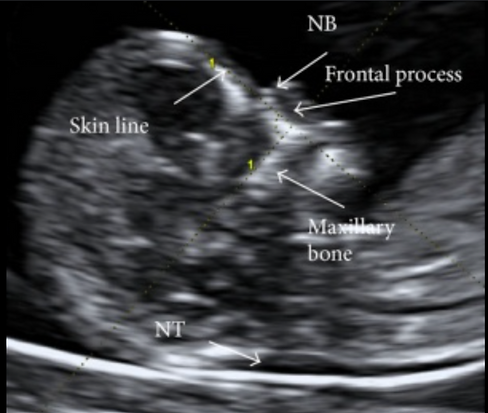
✅ Nuchal Translucency
✅ Placental location and ovaries
With advances in ultrasound technology, your baby's anatomy can be seen in great detail between 12 and 14 weeks gestation. It is now possible to see your baby's arms, legs, fingers, and toes as well as skull, heart, brain, stomach, and kidneys.


Undertaking screening test
During this ultrasound we undertake additional tests to screen your baby (if requested) for , your cervical length, and risks of developing pre-eclampsia. This involves
✅ Chromosomal anomalies by measuring the nuchal tranlucency thickness
✅ Risk of pre-eclampsia by measuring the uterine artery blood flow
✅ Assess for pre-term birth through measuring the length of the cervix
Whether you choose to have screening for chromosomal abnormalities or not, the 1st trimester anatomy scan can provide much more information on your growing baby.
And if you have chosen to undertake NIPT, we welcome you for check of fetal viability on the day of your blood test.


